Minimally invasive surgery VS open surgery
Until the 1980s, traditional open surgical techniques were the only option if surgery was required to treat a patient. Since the emergence of MIS, this has changed. Open surgery tends to require a large incision, lengthy operating times, general anaesthetic, extended recovery periods, and has a greater risk of complications. Visible scarring is also expected after open surgery. MIS offers an attractive alternative. Depending on the procedure, it is usually faster, requires only a small incision or incisions, and can often be done with local anaesthetic. This reduces the risk of complications and shortens the recovery time considerably. It is, therefore, unsurprising that MIS techniques are fast becoming preferable to a more traditional approach.
MIS for breast surgery
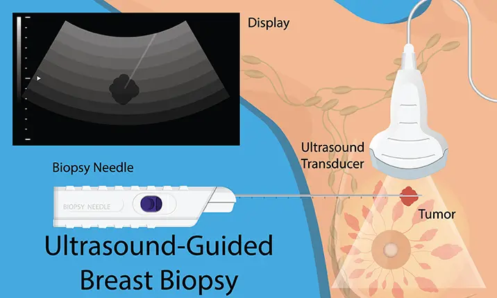
MIS techniques are typically used in the diagnosis of breast cancer. If you find a lump in your breast, and scans and physical examinations are inconclusive, your doctor may need a sample of the affected tissue to run further tests.
This extraction of tissue can be achieved via an ultrasound-guided core needle biopsy for diagnosis, or MIS ultrasound guided vacuum-assisted excision biopsy (removal) of the lump, which is both diagnostic and therapeutic. For suspicious calcifications that are detected on your mammogram, your doctor may recommend stereotactic-guided vacuum-assisted biopsy, which retrieves a good amount of calcification for testing and may also remove all suspicious calcification in the process.
If the biopsy reveals cancer, you will need traditional surgery to remove the mass in its entirety.
MIS techniques include:
- Using image guidance (eg. mammographic or ultrasound guidance) for precision
- Making small incisions that only require 1 – 2 stitches to close, or that do not require any stitches at all
- Core needle and vacuum assisted biopsies that do not require stitches
Why is MIS a preferred option?
This diagnostic procedure results in a better cosmetic result with minimal scarring, hence leading to a more desirable outcome for the patient. In addition, recovery time is often faster.
The importance of self-screening
Cancer develops in stages and early detection plays a vital role in treatment options and survivability. Survival rates are much higher in the early stages, and treatment is less aggressive. If you check your breasts regularly, you will be able to notice any changes and speak to your doctor immediately. A small lump can easily be tested and removed using MIS techniques. Hence, it is important to do self-screening on a regular basis.
How to examine your breasts
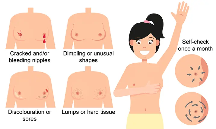
- Look at your breasts in front of a mirror and check for any unusual dimpling, discolouration, or sores
- Examine your nipples and make sure they are not cracked or bleeding
- Check your breasts from all angles, from both sides, the front, and bending forwards, and with your arms raised, looking for any unusual shape
- Lie down and feel your breasts for any lumps or hard tissue, checking each breast and your armpit area. Use flat fingers and press down in circular motions
- Repeat this check once a month after your period, or for menopausal women on a fixed date every month so you won’t forget
What to do if you notice a change
If you find a lump or notice changes in your breasts, refrain from panicking as most lumps are benign. Book an appointment with your doctor as soon as possible in order to get a proper diagnosis. If the cancer is detected in earlier stages, surgical treatment will be less invasive.


