Finding a lump in your breast can be a scary experience and you naturally want to have a diagnosis as soon as possible. Fortunately, around 80% of breast lumps are benign and, with today's modern techniques, the procedure used to make the diagnosis, called a breast biopsy, is usually minimally invasive and can be done in less than half an hour under local anaesthesia. We speak to , a general surgeon specialising in breast surgery, to discover the types of breast biopsies available and what to expect during and after the procedure.
What is a breast biopsy?
A breast lump biopsy obtains tissue from the lump for assessment under a microscope. This is usually done after any unusual breast changes are noticed or if any suspicious or concerning findings are seen on a mammogram or ultrasound. Results from a breast biopsy lab test can be used to diagnose breast cancer and determine the type of treatment to proceed with.
What are the different types of breast biopsies and how are they performed?
There are several types of breast biopsy procedures that are used to obtain a tissue sample. Your doctor will recommend the right one for you based on the size, location and other characteristics of the breast abnormality.
Stereotactic biopsy
This type of biopsy is done using mammograms to pinpoint the location of suspicion areas within the breast.
It is done either by lying face down on a padded biopsy table with one of your breasts positioned in the table or in a seated position. Your breast is firmly compressed between two plates while the suspicious area is located with a targeted mammogram.
The radiologist makes a small incision into your breast and inserts a needle to remove samples of tissue.
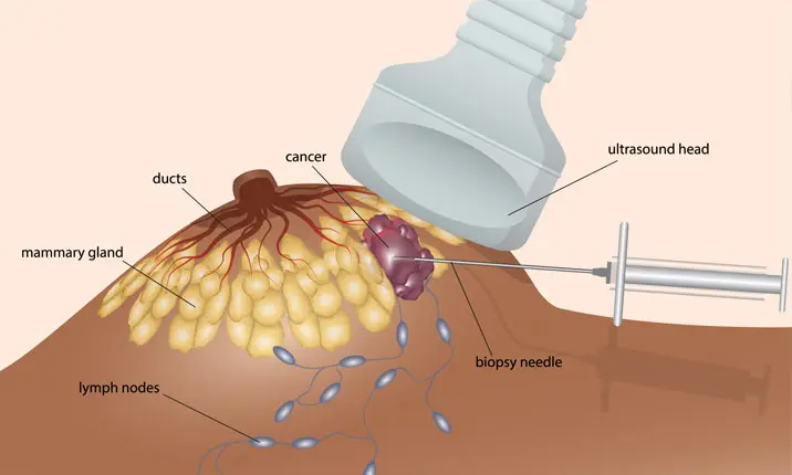
Ultrasound-guided biopsy
Ultrasound imaging is used to guide this type of biopsy.
It is done with you lying down on your back or side on an ultrasound table.
The radiologist locates the mass within your breast using the ultrasound device, makes a small incision and inserts the needle to retrieve several core samples of tissue.
MRI-guided biopsy
This type of biopsy uses MRI imaging, which generates detailed 3D pictures.
During the procedure, you will lie face down on a padded scanning table with your breasts fitted into a hollow depression in the table.
Once the exact location is determined, a small incision is made to insert the core needle and samples of tissue are taken.
What is fine needle aspiration cytology?
This is the simplest type of breast biopsy and may be used to evaluate a lump that can be felt during a breast exam in the clinic, or a lump detected by breast ultrasound.
It is done using a very thin needle that is inserted into the lump. The needle is attached to a syringe that can collect a sample of cells or fluid from the lump.
What is a core biopsy?
In a core biopsy, the breast lump is first located by imaging, then local anaesthesia is injected around the lump to numb the area. Your surgeon will then make a tiny incision in the skin so that a spring-loaded needle can be inserted several times to obtain some tissue from the breast lump. This method has higher accuracy than fine needle aspiration, as it removes more tissue for evaluation.
What is a vacuum-assisted biopsy?
A vacuum-assisted biopsy (VAB) is similar to a core biopsy, but uses a larger needle with a vacuum to pull the tissue into the device. More tissue can be taken in a single insertion and this procedure is often used to remove small breast lumps rather than just taking a sample. Like the core biopsy, a VAB is a minimally invasive procedure, thus causing minimal or no scarring.
What is an open surgical biopsy?
An open surgical biopsy is the conventional way of removing the entire breast lump, and this is done as a day surgery procedure under general anaesthesia. There will be a longer scar, but this procedure allows the most complete removal of larger breast lumps.
Why is a breast biopsy done?
There are several reasons why your doctor may recommend a breast biopsy for you. These may include:
- You or your doctor feel a lump or thickening in your breast during examination, and your doctor suspects breast cancer
- Images from your mammogram show a suspicious area in your breast
- A breast ultrasound scan or MRI reveals a suspicious finding
- Unusual changes are present in your breast, such as crusting, scaling, dimpling skin or a bloody discharge
How do you decide which procedure is best suited for your patients?
It depends on the size and nature of the lumps, and whether there is a decision to remove a lump during the procedure. For tiny lumps (< 5mm) or complex cysts (solid component in fluid), the core biopsy may not be able to obtain good tissue samples while a VAB allows more accurate placement of the needle and better tissue sampling for these lesions. If the lump is causing symptoms of discomfort, and removal is required even if the lump is benign, vacuum-assisted biopsy or open surgical biopsy may be performed.
Does a breast biopsy hurt?
You will feel slight discomfort as the local anaesthesia is administered, similar to having a vaccination. However, you shouldn't feel pain during the biopsy procedure itself. If you do feel pain, you should inform the doctor, who will immediately pause the procedure to administer additional local anaesthesia.
Are there parts of the breast that are more difficult to perform biopsy than others?
Yes, there are. These are typically areas too high up in the armpit, and too close to the chest wall or skin. This is especially true for biopsies done under stereotactic guidance.
It is technically challenging to place the needles in the correct position, or to obtain adequate tissue samples without injuring the chest wall or skin.
In order to evaluate lumps which are hard to reach with stereotactic biopsy, an ultrasound-guided breast biopsy will be preferred. If a breast lump cannot be found using ultrasound, then an open surgical biopsy will be the procedure of choice.
Should women expect some bruising and skin discolouration from the procedure? Are there any general restrictions or home care instructions after a breast biopsy?
You can expect some bruising, but this should resolve in about 1 – 2 weeks. As the breast will be slightly sore in the week following the biopsy, you should refrain from strenuous activity during this period. Mild painkillers will be given and you should take them as required. Let your doctor know if you experience any significant breast swelling.
How soon should a patient expect to receive results of a biopsy?
After your biopsy is done, the breast tissue samples are sent to a lab, where a pathologist examines the sample using specialised tools. The pathologist then prepares a report that is sent to your doctor, who will share the results with you.
The results of a core needle biopsy may take several days to be ready. The results will include details about the size and consistency of the tissue samples, the location of the biopsy site, and whether cancer, non-cancerous (benign) changes or precancerous cells are present. If breast cancer is present, it will include additional information, such as the type of breast cancer you have.
Your doctor will discuss the next steps based on the findings from the biopsy.
How often are breast biopsies positive for cancer?
Only 20% of all breast biopsies are positive for cancer. The remaining 80% of the patients who undergo biopsies do not have cancer.
Is there anything else that you think women need to know about breast biopsies?
Breast biopsies are usually straightforward procedures, performed with minimal risk. How your breast lump is treated thereafter depends on the biopsy results. When the tissue is benign, and the findings are consistent with the imaging, no further treatment is required. However, if cancer cells are found, you will need to undergo further cancer surgery and treatment, even if the entire lump was removed during the biopsy. Another possibility could be that the biopsy result reveals the presence of 'high risk' or atypical cells, which may be associated with surrounding cancer cells. This will usually be deemed inconclusive and you will be required to undergo an open surgical biopsy to get a conclusive diagnosis.


