Cases of skin cancer have been on the rise in recent years, making it the 6th most common cancer in males and the 7th most common cancer among females in Singapore.
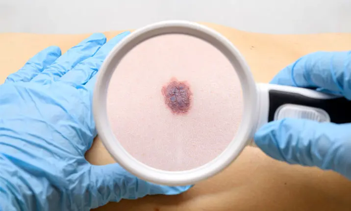
Skin cancers can be broadly classified into melanoma (cancerous mole), which can be potentially life-threatening, or the more common non-melanoma cancers, which are usually caused by long-term sun exposure.
Facing a diagnosis of skin cancer can be overwhelming. To shed light on the treatment options available, dermatologist Dr Tay Liang Kiat shares more about Mohs micrographic surgery, a procedure that can remove skin cancers effectively and precisely.
How are skin cancers usually treated?
In Singapore, non-melanoma skin cancer is more common than melanoma, which is more dangerous with a higher risk of death. Fortunately, most skin cancers are curable when detected early.
There are many treatment options available for non-melanoma skin cancers like basal cell carcinoma (BCC) and squamous cell carcinoma (SCC). These include topical creams, cryosurgery, electrodesiccation and curettage, photodynamic therapy, and oral medications.
A newer surgical procedure, known as Mohs micrographic surgery, is also emerging as a popular treatment option as it can remove cancerous cells in a highly-precise manner while preserving as much healthy tissue as possible.
What is Mohs micrographic surgery?
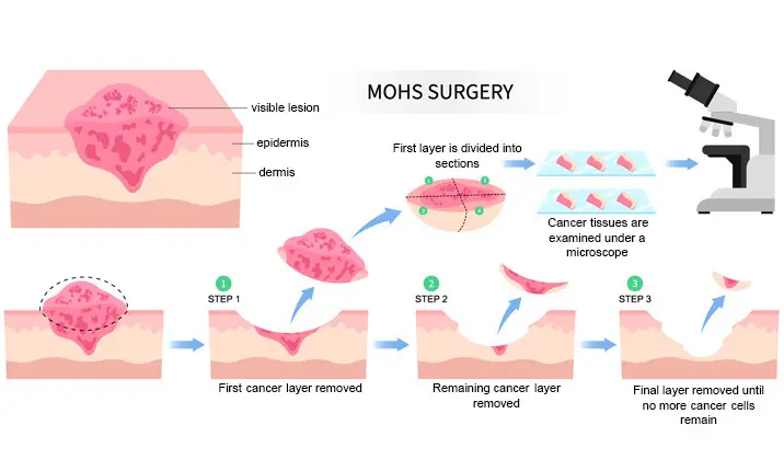
Mohs micrographic surgery is a specialised surgical technique developed by Dr Frederic Mohs in the 1930s to treat skin cancers. The procedure requires the surgeon to methodically cut away thin layers of skin, one layer at a time, and examine each cut tissue section under microscope to ensure that all cancerous cells have been removed.
This is unlike a standard excision, where the surgeon removes the entire tumour together with a border of healthy tissue.
Mohs micrographic surgery is often recommended by doctors for treating high-risk non-melanoma skin cancers. As it is the only treatment modality that examines 100% of the surgical margins, it is effective in removing cancerous lesions with low recurrence rate and provides better cosmetic outcomes as compared to other surgical options.
Mohs surgery is especially useful for removing lesions with these characteristics:
- Larger than 2cm in diameter and found in cosmetically and functionally critical areas, such as the eyes, eyelids, nose, lips, ears, scalp, fingers, toes, or genitals.
- Large and aggressive (spreads quickly).
- Overgrowing with indistinct edges.
- Recurred after previous treatment with radiotherapy, or after incomplete excision, especially for lesions in the head and neck area.
- Diagnosed as aggressive BCC subtypes (infiltrative or micronodular).
- Characterised as SCC with a higher risk of metastasis, usually located on the lip or ears, or in immunosuppressed patients.
Who can perform Mohs micrographic surgery?
Doctors from various surgical fields have tried their hands at Mohs micrographic surgery, but it was only over the past few decades that this surgical technique became fully embraced by dermatologists in many countries. To perform Mohs surgery, a dermatologist would need to have the skill sets of both a surgeon and a pathologist – surgical skills to achieve a clean and precise excision, and a pathologist’s understanding of normal and abnormal tissue structures to differentiate between cancerous and non-cancerous cells.
How is Mohs surgery effective in treating skin cancer?
With Mohs micrographic surgery, the surgeon can thoroughly examine the surgical margins to check for residual cancer cells and ensure that all skin cancer cells are removed. This helps prevent recurrence and allows the maximum amount of healthy tissue to be kept, thus improving recovery outcomes and producing better aesthetic results. In large retrospective studies, Mohs micrographic surgery has been shown to have a curative rate of up to 99% for the first episode of BCC and 97% for the first episode of SCC.
What other types of skin cancer can be treated with Mohs surgery?
Mohs micrographic surgery can also be used to treat other less common skin tumours such as dermatofibrosarcoma protuberans, extramammary Paget disease, and lentigo maligna, among others.
How long does Mohs surgery take?
The time taken depends on the size and location of the tumor. Complex excisions take longer to complete. On average, the procedure may last up to 3 – 4 hours.
How is Mohs micrographic surgery done?
Mohs surgery is done in phases to ensure that all the skin cancer cells are completely removed while keeping the surgical wound as small as possible. Here’s how the procedure is usually performed:
Examination and preparation
Your dermatologic surgeon will examine and clean the area that needs to be treated. Local anesthesia will be injected to numb the area. You will remain awake throughout the procedure.
Top layer removal
In this staged excision phase, the dermatologic surgeon will first cut away a thin layer of visible cancerous tissue to examine the surgical margin.
Sample processing
The removed tissue will be frozen using a cryostat machine and cut into very thin horizontal slices before being stained and mounted on microscope slides.
Lab analysis
The dermatologic surgeon will carefully examine the slides under the microscope to look for cancer cells and map the skin cancer.
Second layer removal
If there are skin cancer cells found at the surgical margins, more tissue will need to be removed. Based on the findings under the microscope and the mapping done earlier, the dermatologic surgeon will remove another layer of skin precisely where the cancer cells were found. This process will be repeated until the skin sample specimen is completely clear of cancer cells.
Reconstruction and wound repair
Once the cancer cells have been completely removed, the wound will be closed with stitches. Smaller wounds in certain areas may be allowed to heal on its own without the need for stitches. Larger wounds may need to be repaired with local skin flap or skin graft to ensure optimal wound healing and for better aesthetic and functional outcomes. The reconstructive surgery is usually performed on the same day as Mohs surgery, once all cancer cells have been removed.
What are the benefits of treating skin cancer with Mohs surgery?
Mohs micrographic surgery offers multiple benefits for patients with skin cancer. These include:
- As the procedure is usually done as a day surgery case, the patient can return home on the same day with minimal downtime.
- The surgery can be performed under local anaesthesia, reducing the risk of general anaesthesia-associated complications.
- The excised samples are tested immediately on-site, which ensures any cancer cells are completely removed before the wound is stitched up. It is unlike standard excision surgery, where the specimen is sent for histology analysis only after the surgery. This eliminates waiting time and the possible need for repeat surgery, thus reducing stress and financial burden on the patient.
- Healthy tissue is preserved as much as possible. Wounds and scars formed after Mohs surgery are usually smaller, heal faster, and have better cosmetic outcomes.
As skin cancer rates continue to rise in the region, it is important to have regular check-ups to help detect any cancers early for successful treatment. If you are looking for an efficient and cost-effective treatment option for skin cancer, speak to a dermatologist to discuss if Mohs micrographic surgery is right for you.


